
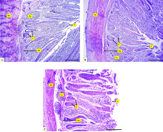
Figure 3:
Microphotographs show the layers of Muscovy duck’s caecum, the muscular layer (Ms), the submucosa (Sm), caecal crypts (C), caecal villi (V), the lymph nodules (Ln) at proximal portion (A), middle portion (B), distal portion (C), Hematoxylin and eosin stain, Scale bar = 200µm.