
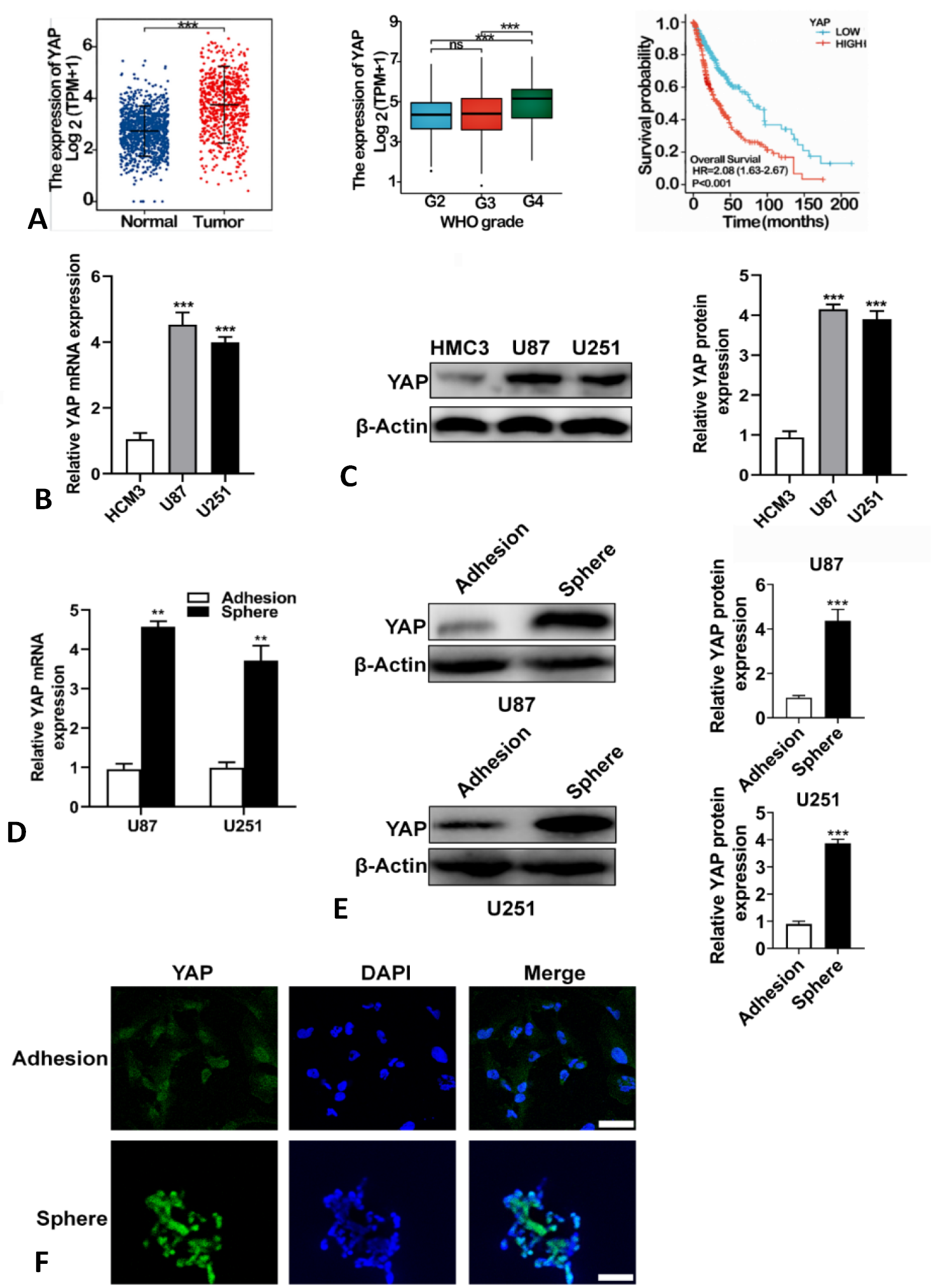
YAP is highly expressed in glioma cells and glioma stem cells. (A) Higher expression of YAP in gliomas (n =689) than in normal brain tissue (n = 1157) was obtained from the TCGA database (***P < 0.0001). Median and interquartile ranges are indicated by black lines. According to WHO grading criteria, (n=635, G2=224, G3=243, G4=168). Medians with interquartile spacing are indicated by black lines. Gliomas (n =697patients) were evaluated with YAP mRNA levels and the results were correlated with an overall survival of 18 years. The red line indicates patients with high YAP transcripts (n = 349) and the blue line indicates patients with low YAP transcripts (n = 348). P values were analyzed by Kaplan-Meier analysis using the GraphPad prism. (B) YAP expression levels were detected in Human Microglia Clone (HCM3) and human glioma cancer cells (U87 and U251) by qRT-PCR. *P < 0.05, **P < 0.01. (C) YAP expression levels were detected in Human Microglia Clone (HCM3) and human glioma cancer cells (U87 and U251) by Western blotting *P < 0.05, **P < 0.01. (D) mRNA expression levels of YAP in spheroid cells and adherent cells. (E) Protein expression of YAP in spheroid cells and adherent cells. (F) Immunofluorescence staining of YAP in spheroid cells and adherent cells in U87 cells (scale bar = 50μm).