
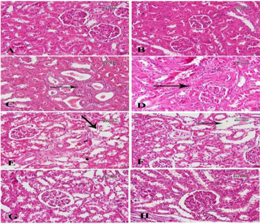
A photomicrograph of sections from renal tissue: (A) From a control -ve group shows the normal structure of the renal glomeruli and the different types of tubules. (B) from a rat treated with corn oil shows no abnormal structure in renal tissue. (C) from a rat treated with bisphenol shows many dilated tubules with necrotic materials within their lumens and increased connective tissue component around them (arrow). (D) Another section for the same group shows massive distortion of renal tissue due to marked increase of connective tissue components (arrow). (F) From a rat treated with bisphenol and Fagonia cretica (low dose) shows slight decrease of fibrosis, but dilated tubules are still noticed (arrow). (F) Another section of the same group shows marked reduction of fibrous, but many tubules show marked dilatation of their lumens with atrophy of lining epithelium (arrow). (G) A section from a rat treated with bisphenol and Fagonia cretica (medium dose) shows many tubules are still dilated with atrophied epithelial lining and no fibrosis around. (H) A section from a rat treated with bisphenol and Fagonia cretica (high dose) shows marked reduction of dilated tubules with no fibrosis.