
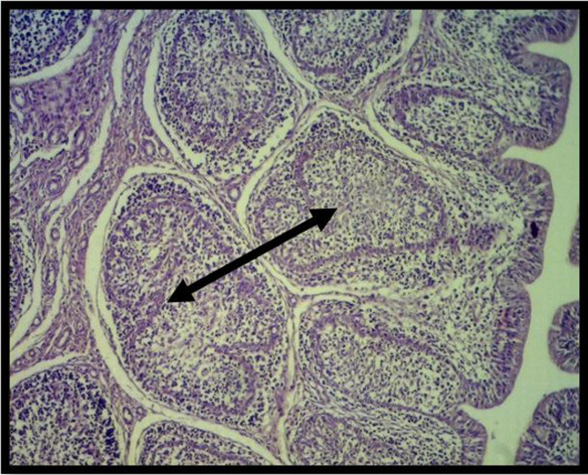
Figure 9:
Histopathological section of testes at 4 weeks of 3rd group shows at various phases of spermatogenesis, seminiferous tubules form, and the Leydig cells and mature spermatozoa fill the lumen. (black arrow) (H&E stainX10).
Histopathological section of testes at 4 weeks of 3rd group shows at various phases of spermatogenesis, seminiferous tubules form, and the Leydig cells and mature spermatozoa fill the lumen. (black arrow) (H&E stainX10).