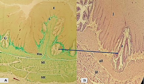
Figure 3:
Photomicrograph section illustrated (A): the esophagus male turkey cervical part the epithelium (E),( lamina propria (L.P) ,musculais mucosa (Mm), two layers of tunica muscularis (MI), (ME) , tunica adventitia (brown arrow), Masson’s Trichrome stain 40× ( B)shows tubular acinar gland in turkey H and E) stain100 ×.