Figure 3:
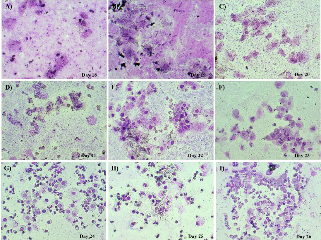
Representative vaginal smear images from non-pregnant pigs during the return estrus stage and subsequent days, illustrating the prevalence of different cell types at various time points.
Representative vaginal smear images from non-pregnant pigs during the return estrus stage and subsequent days, illustrating the prevalence of different cell types at various time points.