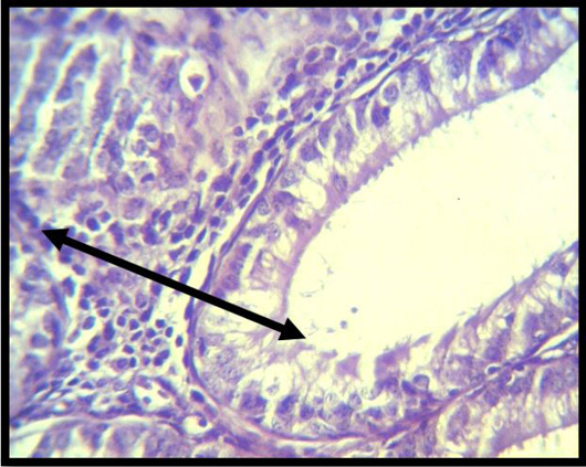
Figure 7:
Histopathological slice of the second group’s testes at 4 weeks shows inflammatory cell aggregation in moderately vacuolated interstial tissue, and the layer of spermatogenic cells is discontinuous. (black arrow) (H & E stain X 40).