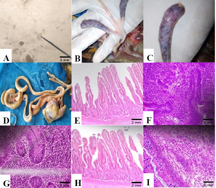
(A) Microscopic examination of protozoal oocyst, (B) Pathological lessions in intestinal parts, (C) Haemorragic and distended intestine (ceca), (D) Nornal intestine and ceca after treatm, (E) Section of lower intestine fed with basal diet showing no haemorrhagic lesion & other changes (microscopic examination 100x, stained with hematoxylin & eosin: H & E), (F) section of lower intestine from infected bird without treatment showing thickening of intestinal lamina propia, presence of coccidial oocysts in lamina propia (microscopic examination 100x, stained with hematoxylin & eosin: H & E), (G) section of lower intestine from infected bird treated with yeast (1g/kg feed) showing mild congestion of sub-mucosal blood vessels, fibrosis & inflammatory infiltration (microscopic examination 100x, stained with hematoxylin & eosin: H & E), (H) section of lower intestine from infected bird treated with yeast (1.5g/kg feed) showing no haemorrhagic lesions, no inflammatory infiltration, sloughing off intestinal villi slightly & no other distinct changes in the intestinal epithelium (microscopic examination 100x, stained with hematoxylin & eosin: H & E), (I) Section of lower intestine from infected bird treated with amprolium showing inflammatory infiltration of intestinal mucosa & congestion of blood vessels (microscopic examination 100x, stained with hematoxylin & eosin: H & E).