
Fig. 2.
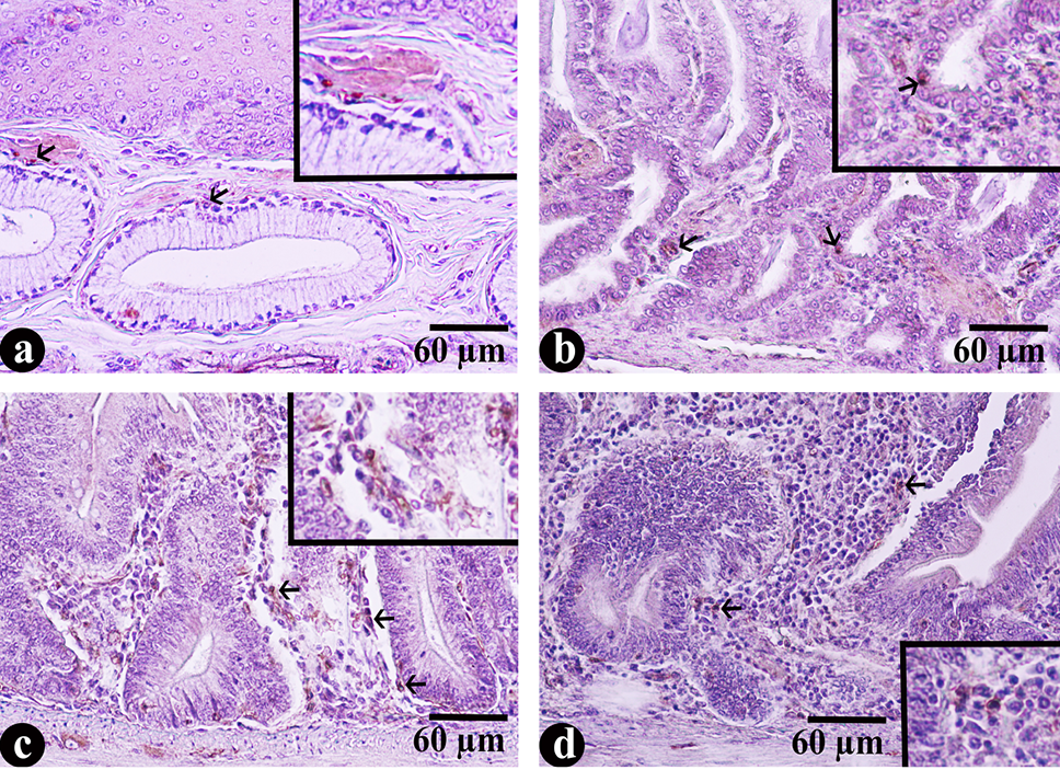
Histological structure of gastrointestinal tissues of red legged partridge stained with anti-VIP antibodies; (a) proventriculus; (b) ventriculus; (c) duodenum and (d) jejenum. Arrows show immunoperoxidase positive stained cells.
Histological structure of gastrointestinal tissues of red legged partridge stained with anti-VIP antibodies; (a) proventriculus; (b) ventriculus; (c) duodenum and (d) jejenum. Arrows show immunoperoxidase positive stained cells.