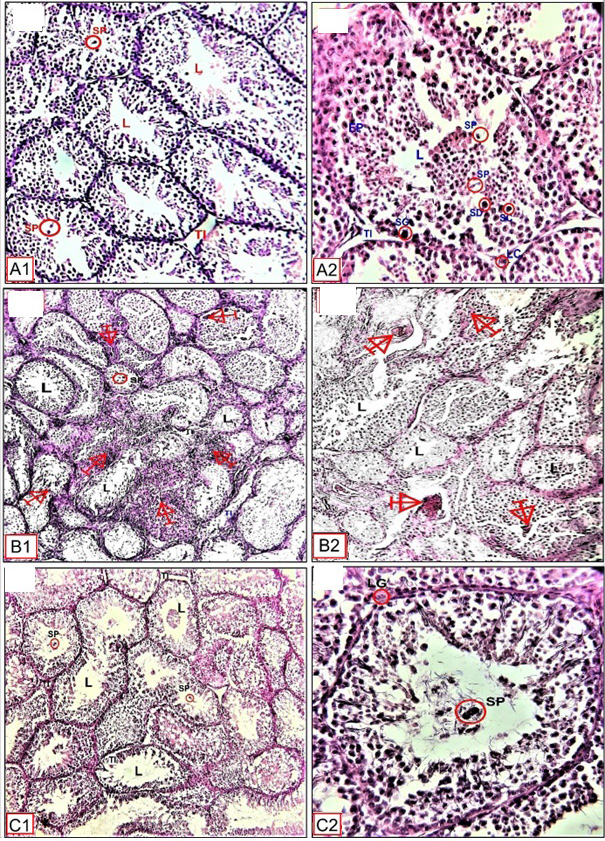
Fig. 5.
Histological structure of testis control, SPT, and TT100 treated pigeons under a long photoperiod ((19L:5D) showing: control group (A1 and A2) normal epithelial tissue (EP), Lumen (L), Leydig cell (LC), Sertoli cell, Interstitial tissue (TI), Spermatozoa (SP), Spermatogonia (SG), Spermatocyte (SC), Spermatid (SD). SPT group (B1 and B2) degenerative in the testicular tissue and irregular seminiferous tubules are observed. TT100 groups (C1 and C2) showed normal testicular tissue and increased spermatozoa and epithelial germ cells number. Magnification: A1, B1, C1= X10. A2, B2, C2= X40. Stain H and E.