
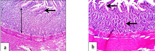
Figure 6:
Representative photomicrograph of gander intestine in G3 showing focal apical villus sloughing (arrow) with mild cellular infiltrates and mild to moderate crypt proliferation (a) and intactness of most intestinal architecture (b). (H & E, x100).