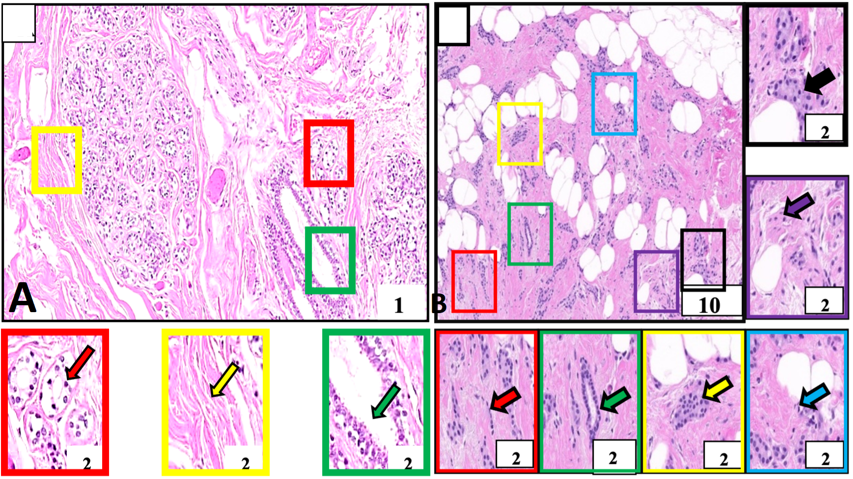
Histological structure of normal and breast tumor samples of our study subject identified for invasive ductal carcinoma. A, Negative control tissue showing morphologically normal breast cells. These cells and their nuclei are smaller, uniform, and regular in size and shape (red outlined box and arrow). The breast stroma is normal and does not show cellular penetration (yellow outlined box and arrow). The milk-carrying ducts are also nicely lined with normal cell layers (green outlined box and arrow). B, Breast cancer tissue. This tissue is showing malignant cells (red outlined box and arrow) identified in milk-carrying ducts of the breast. These cells and their nuclei are larger in size and pleomorphic morphologically (purple outlined box) arranged in cords (red outlined box and arrow), nests (yellow outlined box and arrow), sheets (black outlined box and arrow), forming tubular structures (green outlined box and arrow) and showing mitotic figures (blue outlined box and arrow). These cells are penetrating the rounding breast stroma (purple outlined box and arrow). Boxes with various color outlines are showing digitally magnified corresponding selected areas on slides A and B. Stain: Hematoxylin and Eosin.