
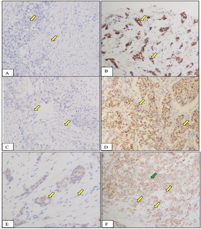
Expression of ER (A, B), PR (C, D), and HER2/neu (E,F) receptor (protein) in normal and tumor tissues from female breast cancer patients recruited in this study. A, C, E, Negative control tissue: cells in this tissue are not showing any nuclear stain due to the absence of specific receptors in these cells. On the other hand, they are showing blue color staining because of H&E counter stain. B, D, F, Breast cancer tissue, nucleic of cancer cells are showing intense staining (yellow arrow) due to the presence of overexpressed specific receptors. These receptors are localized in the cell nuclei and upon application of specific for each antibodies to these receptors, they produce a brown color. Stain: Immunohistochemical stain specific to ER receptor; Magnification: 20X; Counterstain: Hematoxylin and Eosin, Magnification: 20X.