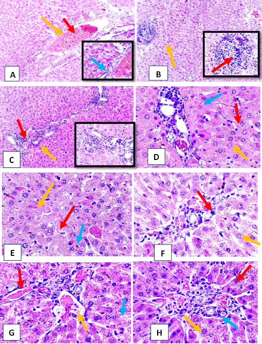
Histopathological section of liver tissue from tetrodotoxin treated group (A, B, C, D, E) (0.5 µg/kg b.w.) of male albino rats (A) obvious dilation and congestion of portal vein (red arrow) with mild cellular infiltration (yellow arrow) with evidence of scattered apoptotic hepatic cells (blue arrow). (B) moderate vascular congestion and dilation of hepatic vessels (red arrow) with focal aggregation of mononuclear cells formation granulomatous like lesion (yellow arrow). (C) moderate periductal mononuclear cells aggregation (red arrow) with slight presence of edematous substance in adjacent hepatic parenchyma (yellow arrow). (D) various form of nuclear necrosis (red arrow) with number apoptotic hepatocyte (yellow arrow) with periductal mononuclear cells infiltration (blue arrow). (E) moderate nuclear pyknosis of hepatocyte (red arrow) with slight sinusoid congestion (yellow arrow) with evidence of councilman body (blue arrow). (F, G, H) (1 µg/kg b.w) (F) perivascular and periductal mononuclear cells infiltration (red arrow) with moderate necrotic finding of surrounding hepatocyte (yellow arrow) (G) hepatocyte vessels dilation and congestion (red arrow) with new neutrophil in lumen (yellow arrow) with binucleted hepatocyte (blue arrow) (H) moderate perivascular mononuclear cells (res arrow) with numerous binucleated hepatocyte (yellow arrow) with scattered apoptotic cells (blue arrow) (H&E stain, X40).