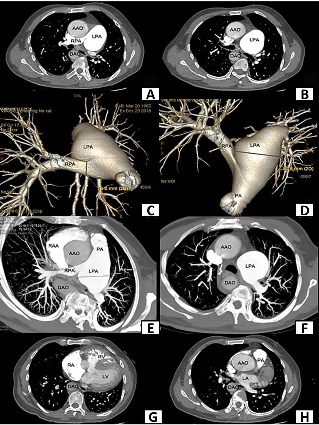
Enhanced cardiac computed tomography(CT) scan before surgery shows aortic overriding, ventricular septal defect, RVH and obstruction of right ventricular outflow tract (AAO, ascending aorta; DAO, descending aorta; LPA, left pulmonary artery; RPA, right pulmonary artery; PA, pulmonary artery; RAA, right atrial appendage; RA, right atrium; LA, left atrium; RV, right ventricle; LV, left ventricle). A, CT shows aortic override on the right pulmonary artery trunk and the dilated left pulmonary artery. B, CT shows dilated left pulmonary artery. C, CT shows stenosis of right pulmonary artery trunk and dilated left pulmonary. D, CT shows stenosis of right pulmonary artery trunk and dilated left pulmonary. E, CT shows aortic overriding, stenosis of right pulmonary artery trunk and dilated left pulmonary artery. F, CT shows dilated left pulmonary artery and thickened pulmonary branches. G, CT shows right ventricle hypertrophy and ventricular septal defect. H, CT shows aortic overriding and obstruction of right ventricular outflow tract.