
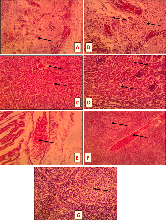
Tissue sections of S. aureus and /or E. coli infected birds at 3 DPI (HandEX, 200) showing the following lesions. A: Subcutis of E. coli infected bird: Severe suppurative inflammation characterized by heterophils and mononuclear cells infiltration in s.c tissues (head of arrow). B: Subcutis of S. aureus and E. coli infected bird: severe suppurative inflammation with marked accumulation of heterophils and mononuclear cells in s.c fatty tissue (head of arrow). C: Liver of S. aureus infected bird: hydropic degeneration of the hepatocytes (head of arrow). D: Kidney of S. aureus + E. coli infected bird: severe hydropic degeneration (head of arrow). E: Subcutis of S. aureus infected bird: subcutaneous hemorrhages (head of arrow). F: Spleen of S. aureus infected bird: congested red bulb and blood vessels with necrotic area (head of arrow). G: Bursa of E. coli and/or S. aureus infected bird: depletion of the lymphoid follicle (head of arrow).