
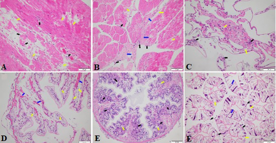
Histopathological observation of various organs in diseased P. clarkia. A-B: The diseased crayfish muscle tissue showed inflammatory cell infiltration (black arrow), cell necrosis (yellow arrow) and fibrinoid degeneration (blue arrow). C: A large number of inflammatory cells infiltrate into the gill fila of the diseased crayfish (black arrow), vacuolar degeneration (yellow arrow). D-E: A large number of inflammatory cells infiltrate the intestines of the diseased crayfish (black arrow), showing vacuolar (yellow arrow) and hyaline degeneration (blue arrow). F: The lumen of the hepatic tubule dilates, the star-shaped structure disappears (the blue arrow), hepatocyte structure collapse, vacuolization (the yellow arrow), interstitial inflammatory cell infiltration (the black arrow).