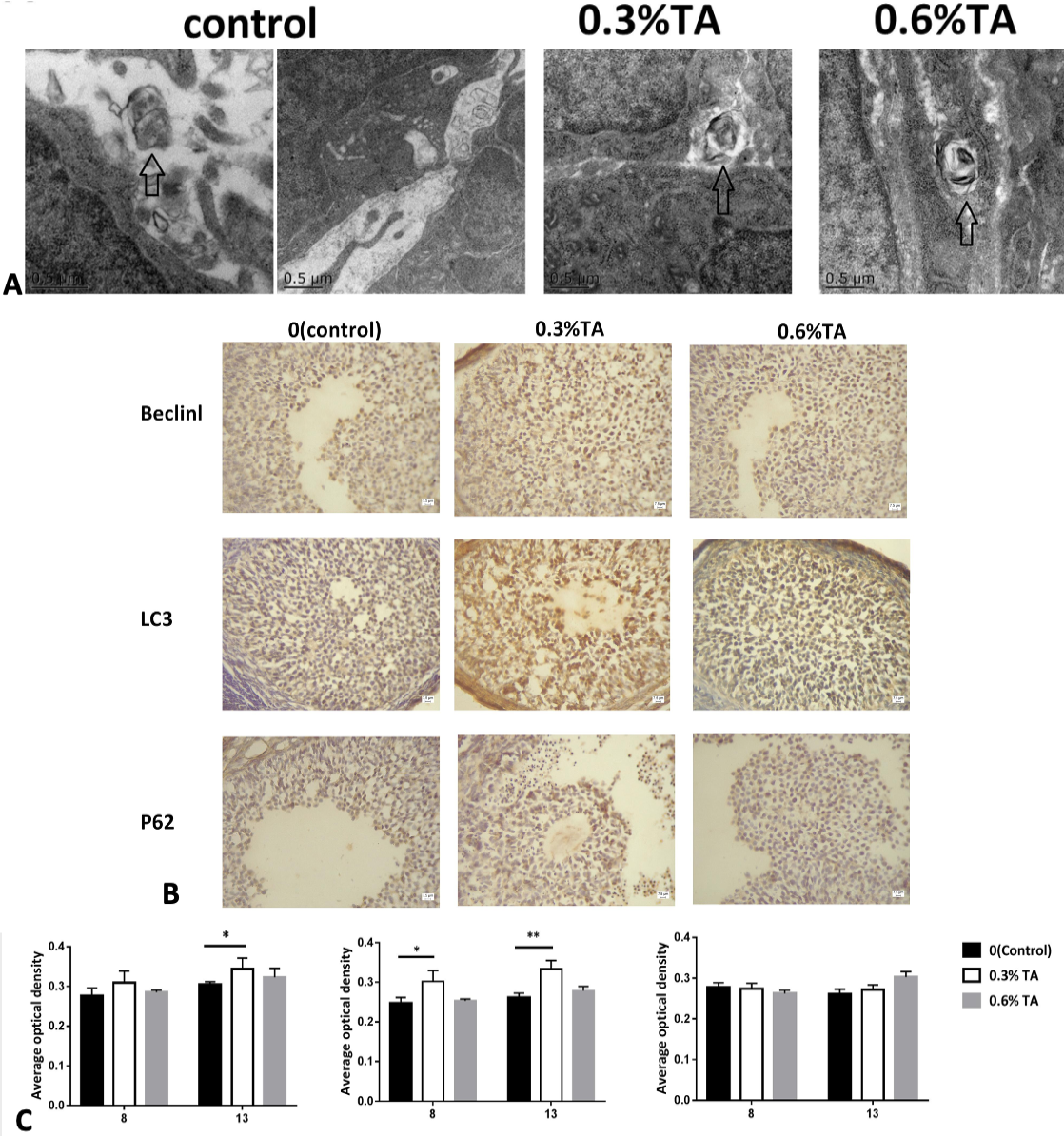
(A) Electron microscopy of the ovaries treated with different doses of tannic acids. The arrows represent autophagosomes. (B) Immunohistochemical staining of Beclin1 protein in the ovary. The immunostaining of the ovaries of the control animals was weaker. The ovaries in the adult low-dose group had stronger immunostaining. Immunohistochemical staining of LC3 protein in the ovary. The immunostaining of the ovaries of the control animals was weaker. Strong immunostaining of the ovaries in the low-dose and high-dose groups. In the ovaries, granulosa cells are strongly stained. Immunohistochemical staining of P62 protein in the ovary. There was no significant difference in immunostaining in each treatment group. (C) The effect of different doses of tannic acid on Beclinl, LC3 and P62 proteins during adolescence and sexual maturity (n=8). Data are expressed as the mean ± sem. * P < 0.05., **P < 0.01
