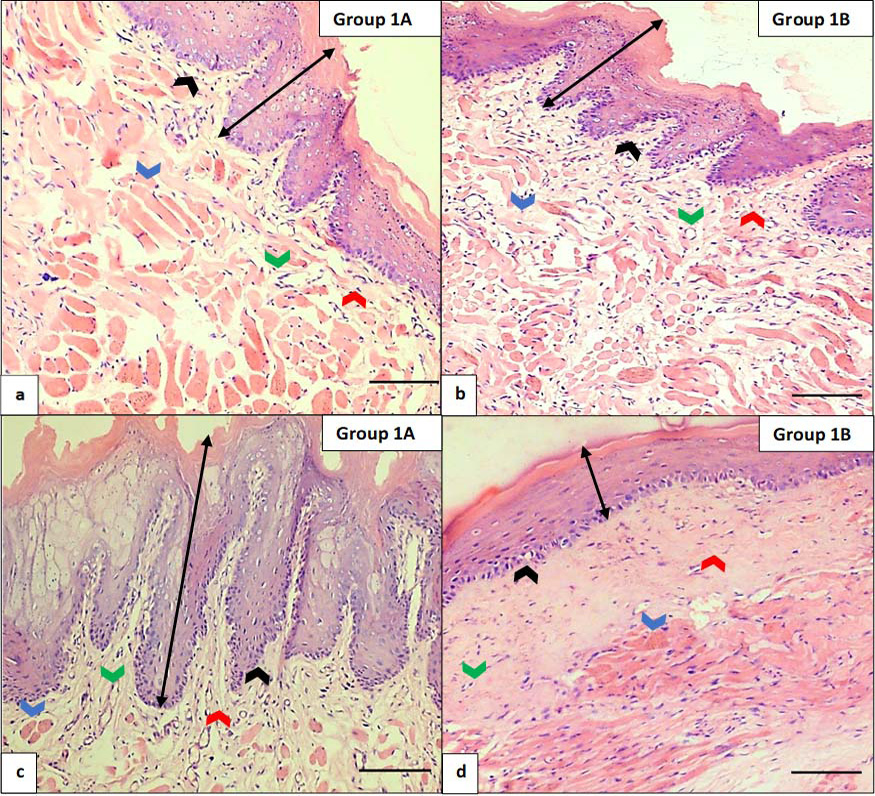
Fig. 2.
Effect of bleomycin on histological structure of buccal mucosa of rat after treatment for 4 weeks and 8 weeks. (a) Control group 1A at week 4 (scale bar=100 µm). (b) Experimental group 1B at week 4 (scale bar=100 µm). (c) Control group 1A at week 8 (scale bar=100 µm). (d) Experimental group 1B at week 8 (scale bar=100 µm). In the 4th week, the control group 1A (a) showed normal buccal mucosa features i.e., thickened stratified squamous epithelium with orthokeratosis (double black arrows) having long and narrow rete pegs (black arrows), moderate number of vessels (green arrow), absence of inflammatory infiltrate, normal amount of collagen in connective tissue (red arrows) and muscle fibers in the form of fascicles (blue arrows). In the 4th week, the experimental group 1B (b) showed early stage OSMF features i.e., slight decreased thickness of epithelium with same degree of orthokeratosis (double black arrow), shortened rete pegs (black arrow), few constricted vessels and inflammatory infiltrate, pink filaments in connective tissue just below the epithelium (red arrow) and no muscular atrophy (blue arrows). In the 8th week, the control group 1A (c) showed normal buccal mucosa features i.e., thickened stratified squamous epithelium with orthokeratosis (double black arrows) having long and narrow rete pegs (black arrows), moderate number of vessels (green arrow), absence of inflammatory infiltrate, normal amount of collagen in connective tissue (red arrows) and muscle fibers in the form of fascicles (blue arrows). In the 8th week, the experimental group 1B (d) showed advanced stage OSMF features i.e., epithelial atrophy with increased orthokeratosis (double black arrows), absent rete pegs (black arrows), constricted and absent vasculature (green arrow), reduced cellularity, hyalinized change in collagen in connective tissue (red arrows) and muscular atrophy (blue arrows) (Stain: hematoxylin and eosin, Magnification: 10X).