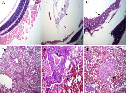
Histopathological lesions in the trachea and lungs of naturally infected quails: (A) trachea of normal quail (non-infected) showing normal wall and lumen, (HE, x100); (B) trachea of infected quail with NDV showing mucus exudate inside tracheal lumen(arrow), (HE, x100); (C) trachea of infected quail with NDV showing thickened tracheal wall by leukocyte(arrow) and proliferative glands (arrow head), (HE, x400); (D) lung of normal quail showing normal pulmonary tissue, (HE, x100); (E) lung of infected quail with NDV showing bronchial exudate (arrow), necrosed muscles (arrow head) and sero fibrinous fluid in adjacent air vesicle, (HE, x400);(F) lung of infected quail with NDV showing focal pneumonic area fibroblasts, macrophages and giant cells within pulmonary exudate (arrow), (HE, x100).