
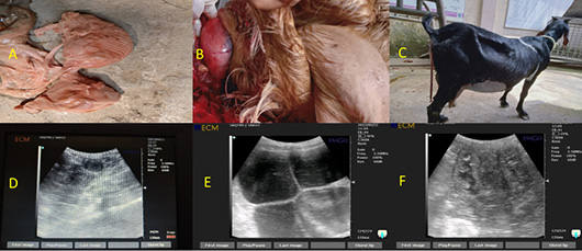
Figure 1:
(A) An aborted foetus of 3.5 months gestational age; (B) A ball-like, round structure protruding from the vulva indicates prolapse of the vagina; (C) Hanging the placenta from the vulva of the doe after parturition indicates the retention of the placenta; (D) A round hypoechoic image within the ovary on ultrasonography indicates a luteal cyst of the ovary; (E) An echoic sac within the uterus on ultrasonography indicates a fluid-filled condition in the uterus, known as hydrometra; (F) A hypoechoic sac in the uterus indicates mucous present within the uterus, known as mucometra.