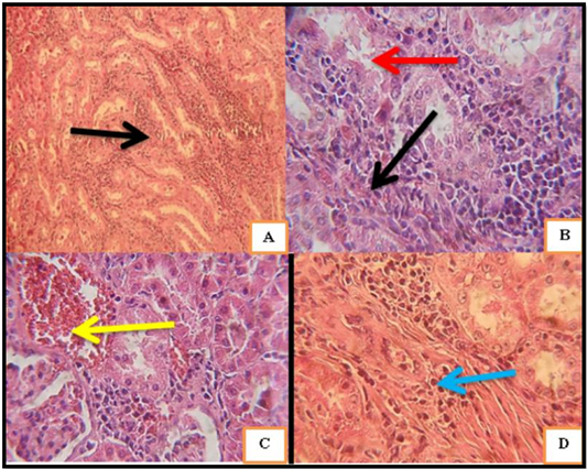
A: (10X H and E) sections of kidney from male rats (control group) showed the normal renal glomerulus (black arrow) and showed the normal renal tubules. B: sections of kidney (T. terrestris extract group), showed focal interstitial nephritis appears as aggregations of inflammatory cells (Black arrow) some tubules appears loosen its lumen and desquamated epithelial lining due to necrosis (Red arrow). C: Sections of kidney treated with (lead acetate group) showed hyaline casts appears with hemorrhagic changes enclosed to focal aggregation of monocytes (Yellow arrow), (40X H and E). D: Sections of kidney treated with (lead acetate + T. terrestris extract group) fibrosis appears very clear near to inflammatory cells foci which reveals presence of chronic inflammation (Blue arrow), (40X H and E).
