
Figure 4:
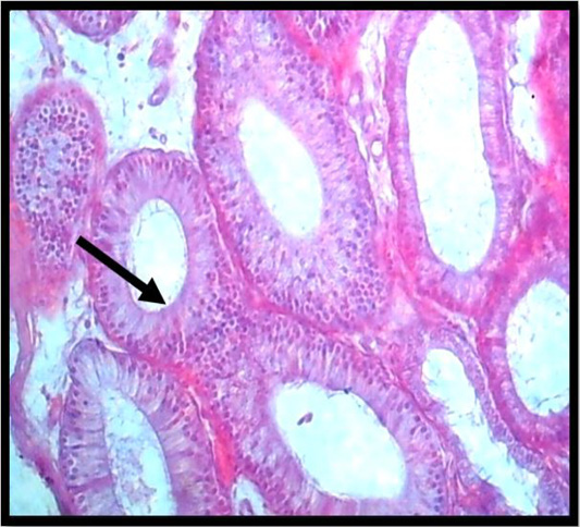
Histopathological slice of the first group’s epididymis at 4 weeks demonstrates granuloma development (black arrow) (H & E stain X 40).
Histopathological slice of the first group’s epididymis at 4 weeks demonstrates granuloma development (black arrow) (H & E stain X 40).