
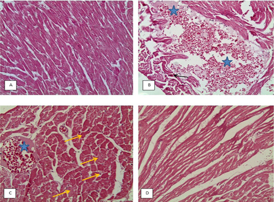
Figure 6:
(H&E X20), A: heart of CG, B: heart of TG showing large area of hemorrhage and edema, myocardiolysis (star) and necrosis of muscle (black arrow), C: heart of DAG showing myocardiolysis, hemorrhage and edema (star), swelling of muscles and muscle necrosis (yellow arrow), D: heart of BG more or less to normal with slight myocardiolysis.