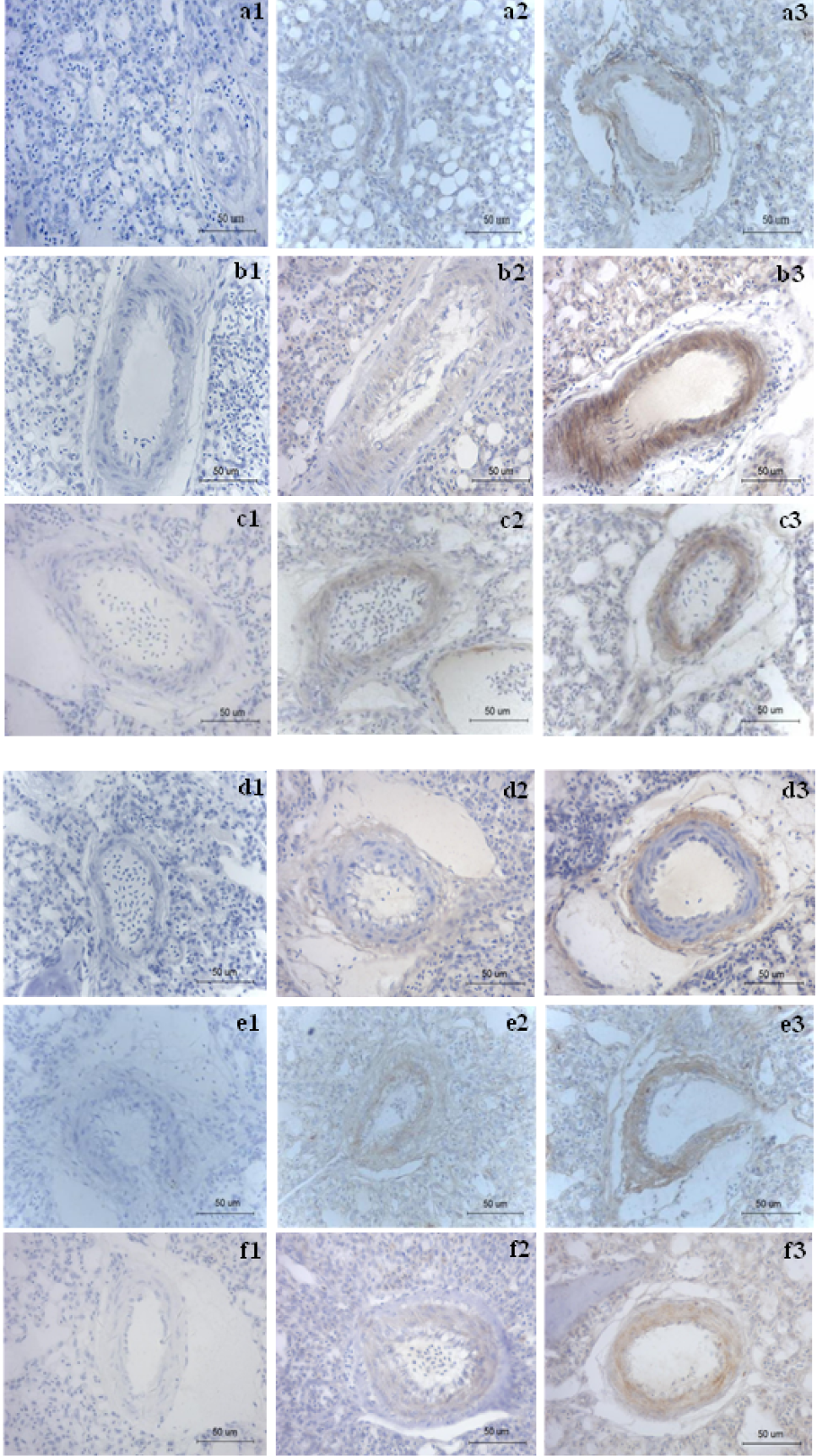
Fig. 3.
Immunohistochemical analysis of protein in broiler lungs (magnification: 400×). a, MIF; b, p-Raf; c, p-ERK; d, p-Akt; e, p-GSK3β; f, cyclin D1; 1, negative control with PBS; 2, the lungs of healthy broilers; 3, the lungs of PH broilers.