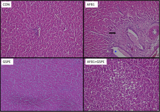
Figure 3:
Photomicrographs of liver sections from quails in the different groups: Con, control quails showing normal hepatocytes arranged in plates radiating from the central vein and separated by blood sinusoids; AFB1, quails treated with aflatoxins alone showing marked fibrosis of portal area (→) associated with focal dilation of hepatic sinusoids; GSPE, quails treated with GSPE revealing normal hepatocytes and D, quails treated with (AF+GSPE) showing large intra-hepatic fat droplets.