Figure 4
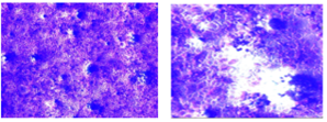
Microscopic images show morphological alteration of AMN3 cell lines. (left) treated with AGE, (right) untreated cells. The cells were stained with crystal violet (20X).
Microscopic images show morphological alteration of AMN3 cell lines. (left) treated with AGE, (right) untreated cells. The cells were stained with crystal violet (20X).