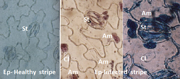
Figure 5:
Photograph showing the crystal (Cl) and amorphous (Am) inclusion bodies in cells of epidermal (Ep) strips and stomata (St) of infected and healthy N. glutinosa leaves.
Photograph showing the crystal (Cl) and amorphous (Am) inclusion bodies in cells of epidermal (Ep) strips and stomata (St) of infected and healthy N. glutinosa leaves.