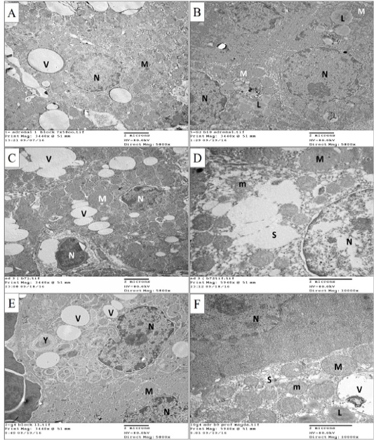
Electron microscopic pictures for the zona fasciculata cells of the adrenal cortex from different groups. (A) Cells of the control group showing rounded regular nucleus (N) with dispersed chromatin, multiple vesicular mitochondria (M) and vacuoles (V). (B) Cells of the control exercise group, showing rounded nuclei (N) with dispersed chromatin, multiple vesicular mitochondria (M). Notice the multiple vacuoles with lipid content (L). (C) Cells of the depression group showing shrunken irregular nuclei (N), swollen mitochondria (M) and multiple vacuoles of different sizes (V). (D) Cells of the depression group showing some swollen mitochondria (M), mitochondria with destructed cristae (m) and dilated vesicular smooth endoplasmic reticulum (S). (E) Cells of the depression exercise group showing normal sized nuclei (N) with irregular contour and multiple normal vesicular mitochondria (M). Few vacuoles (V) and lysosomes containing substance (Y) appear in the cytoplasm. (F) Cell from the depression exercise group showing normal nucleus (N), mitochondria with normal size and cristae (m) and nearly normal smooth endoplasmic reticulum (S). Few mitochondria appear dilated (M). Few vacuoles (V) appear with some showing lipid content (L). A, B, C and E x5800, and D and F x10000 - scale bar 2µ.