
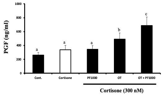
Figure 3:
Effects of cortisone on PGF production by cultured bovine endometrial tissue at follicular stage (Mean ± SEM, n=4 experiments, each performed in triplicate). Endometrial tissues were exposed to cortisone (300 nM) or cortisone (300 nM) combined with oxytocin (OT, 100 nM) in the presence or absence of PF (1000 nM; PF 1000) for 4 h. Different superscript letters indicate significant difference (p<0.05) as determined by ANOVA followed by protected least significant difference test (PLSD).