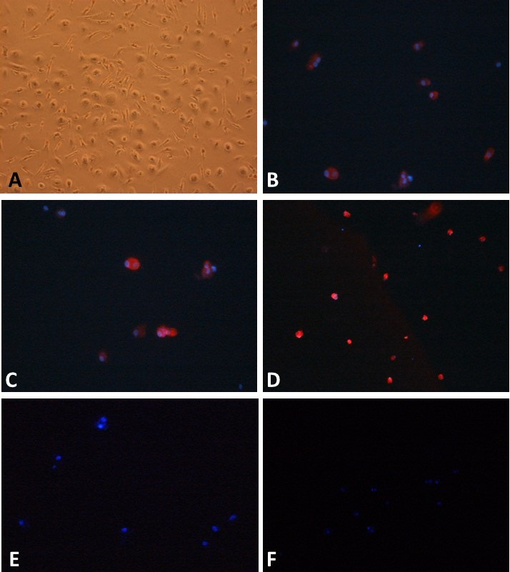
Cultured human CD34+ erythroid progenitor cells (EPCs). A, cells in erythroid expansion supplement medium. Numerous mononuclear cells having regular complete cell membrane and round nuclei are visible under inverted microscope. B, shows bright fluorescence emitting after binding with the corresponding antibody as observed under fluorescence microscope. C shows CD71+ EPCs as bright fluorescent cells after binding with the corresponding antibody when labeled with anti-human CD71 PE antibody observed under fluorescence microscope. D, CD235a+ EPCs are identified as bright fluorescent cells after binding with corresponding antibody. Observed under fluorescence microscope, when labeled with anti-human CD235a PE antibody. E, EPCs did not emit fluorescence signal as they did not bind to anti-human CD3 antibody when labeled with anti-human CD3 PE antibody. This also excludes the contamination with CD3+ cells. Cells were counter stained with DAPI (showing blue fluorescence). Cells were observed under fluorescence microscope. F, EPCs did not emit fluorescence signal as they did not bind to anti-human CD14 antibody when labeled with anti-human CD14 PE antibody. This also excludes the contamination with CD14+ cells. Cells were counter stained with DAPI (showing blue fluorescence). Cells were observed under fluorescence microscope. Magnification: 20 X.