
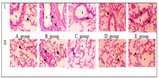
Figure 5
The effect of famotidine on the lung tissue histopathology. The process of OVA-sensitization resulted in a noticeable increase in the accumulation of leukocytes in lung tissue. However, in rats treated with famotidine, this accumulation was reduced, highlighting the anti-inflammatory effect of famotidine. Lung tissue photos were taken by light microscope, X-40, H&E stain. The arrows point to inflammatory cells; b, bronchioles; a, alveoli; A_group, negative control; B_group, positive control; C_group, prednisolone; D_group, famotidine; E_group, prednisolone and famotidine.