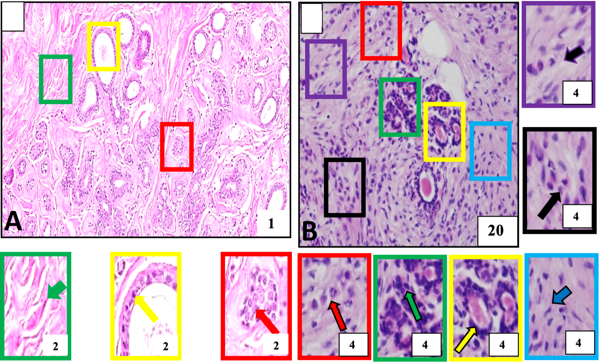
Histological structure of normal and breast tumor samples of our study subject identified for invasive lobular carcinoma A, Negative control tissue showing morphologically normal breast cells. These cells and their nuclei are smaller, uniform, and regular in size and shape (red outlined box and arrow). The breast stroma is normal and does not show cellular penetration (green outlined box and arrow) and normal cell layers constituting breast milk carrying ducts (yellow outlined box and arrow). B, Breast cancer. The tumor tissue is showing a population of malignant cells across the tissue and is detected the in lobular part of the breast. These are larger than normal breast cells arranged into single files or chains of cells (red outlined box and arrow), single cells (purple outlined box and arrow), cells with evident cellular and nuclear pleomorphism (black outlined box and arrow), round to oval shaped nuclei (green outlined box and arrow) aggregate around ducts (yellow outlined box and arrow) and penetrating surrounding breast stroma (blue outlined box with arrow). Boxes with various color outlines are showing digitally magnified corresponding selected areas on slides A and B. Stain: Hematoxylin and Eosin.