
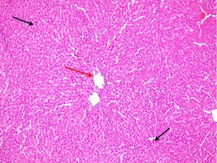
Figure 2:
A photomicrograph of a control group rat liver tissue segment revealed hepatic tissue with normal liver lobules (black arrow) and a remarkable normal central vein (red arrow). (10X H and E).
A photomicrograph of a control group rat liver tissue segment revealed hepatic tissue with normal liver lobules (black arrow) and a remarkable normal central vein (red arrow). (10X H and E).