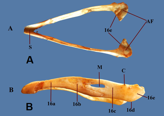
Figure 6:
photograph showing the mandible. A: Dorsal view; B: Lateral view.
1-Occipital bone, 1a-Supraoocipital part, 1b-Basioociptal part, 1c- Exo-oxcipital part, 1d-Occipital condoyle, 2-Parital bone, 3-Frontal bone, 3a- longitudinal fissure, 4- Sphenoid bone, 4a-Presphenoid, 4b-Basisphenoid, 5-Squamus part of temporal bone, 5a-orbital process, 5b-Zygomatic process, 5c-temporal fossa, 6- Ear capsule, 6a- Internal acoustic meatus, 6b- Tympanic cavity proper, 7- Lacrimal bone, 7a- dorsal lacrimal process, 7b-ventral lacrimal process, 7c- deep fissure, 8-Quadrate bone, 8a- Orbital process, 8b- Otic process, 8c- Articular process, 9- Zygomatic (Jugal) bone, 9a Jugal process , 9b- Proper jugal, 9c- Quadrojugal process,10- Palatine bone, 11-Vomer bone, 12- Nasal bone, 12a-Frontal process of Nasal, 12b- Maxillary process of Nasal, 12c- Premaxillary process of Nasal, 12d- Frontonasal hing 13- Inscisive bone (premaxillary bone), 13a- Frontal process of incisive, 13b- Maxillary process of incisive , 13c- Palatine process of incisive, 14-Maxillary bone, 14a-Zygomatic process, 14b- Palatine process of maxilla, 15-Ethmoid bone, 15a –vertical part of Ethmoid, 15b- Horizontal part of Ethmoid, 16- Mandible, 16a- Dental bone, 16b-splenial bone, 16c- supra angular bone, 16d-The angular bone, 16e-The articular bone. N: Nuchal crest, FM: Foramen Magnum, OP: Optic foramen, E: Ethmoid foramen, PF: Palatine fissure, PT: Pterygoid facet, M: Mandibular foramen, C: Coronoid process, S: Mandibular Symphysis, AF: Articular facet for quadrate bone, OL: olfactory foramen.OC: Orbital Cavity, ON: Osseous opening of nostril, NS: Nasal Septum, CC: Cranial cavity.