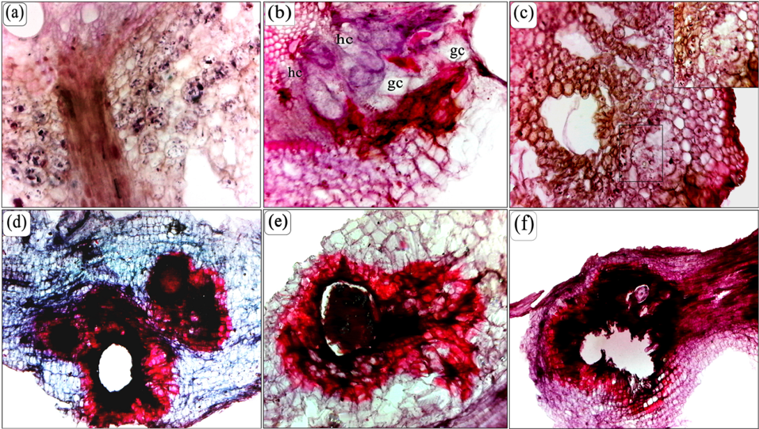
(a) Transverse section of a banana root showing the granular structures in the cells of cortex layer near the feeding site of M. incognita. (b) Note the nuclei in the vicinity of nematodes either enlarged or disintegrating and either cytoplasm contracting from cell walls still intact or contracted and cell walls beginning to rupture. In addition, giant cell transforming from undifferentiated hypertrophied cells. (c) Transverse section through the cortex noted the extensive cavity in the cortex, the nematodes at the periphery of the lesion and some cell fragments towards the center necrotic and lignified cells. (d, e and f) Longitudinal section of a banana root through the gall showing the laceration and deformation area and phenolic and lignified cells of damaged cortex extended towards the pericycle on the old root and the root hair. (Scale bars: a-f = 200 μm).