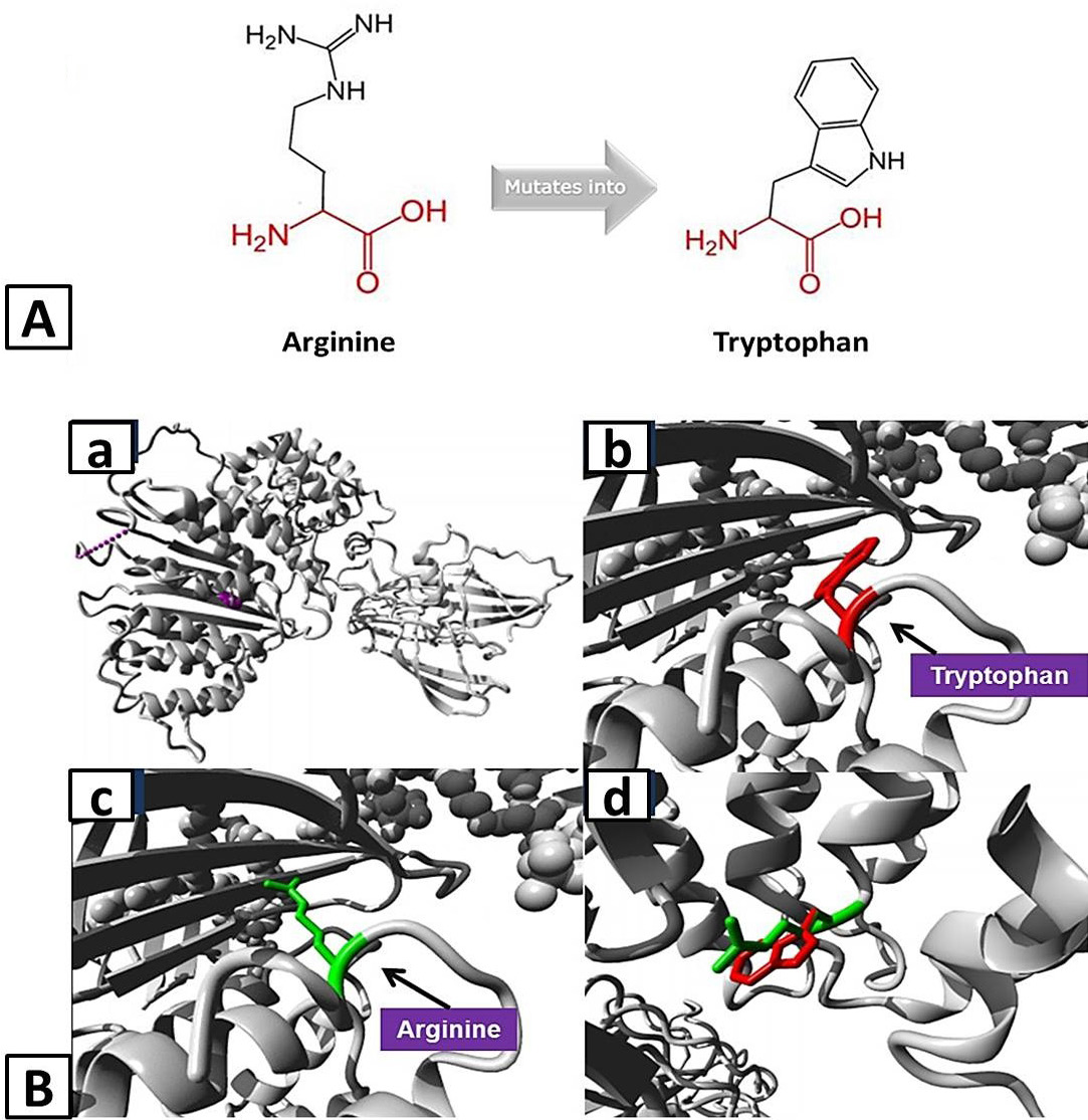
A, Schematic structures of the original (left) Arginine and the mutant (right) amino acid tryptophan for variant Arg532Trp of APOB created by HOPE tool. The backbone, which is the same for each amino acid, is colored red and the side chain, unique for each amino acid, is colored black. B, Homology models of ApoB representing structural impact of variant Arg532Trp: a, Overview of the protein in ribbon-presentation with protein is colored grey, and the side chain of the mutated residue is colored magenta shown as small balls; b, Close-up of the mutation with protein is colored grey and red represent side chain of mutant residue; c, Close-up of the mutation with protein is colored grey and green represent side chain of wild-type residue; d, Close-up of the mutation with both wild-type and mutant residues side chain on the protein.