
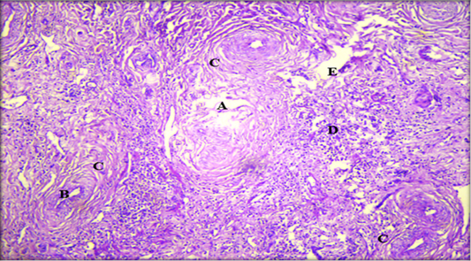
Figure 5:
Histopathological section of liver showing; A: destruction of liver tissue; B: thickening of bile duct; C: fibrosis proliferation in the portal area; D: coagulative necrosis of liver cell; E: parasitic tract formed from destructed hepatocytes intermixed with infiltrated inflammatory cell (H & E stain. X10).