Figure 6:
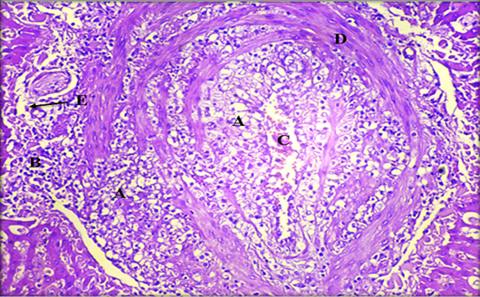
Histopathological section of liver showing; A: fatty change; B: coagulative necrosis; C: hemorrhage; D: fibrous connective tissue capsule; E: Kupffer cells (H & E stain. X20).
Histopathological section of liver showing; A: fatty change; B: coagulative necrosis; C: hemorrhage; D: fibrous connective tissue capsule; E: Kupffer cells (H & E stain. X20).