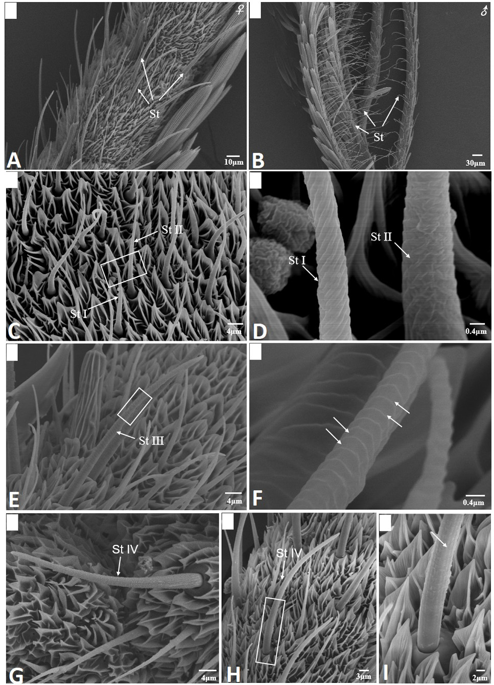
Fig. 2.
Scanning electron micrographs of sensilla trichodea in Scopula subpunctaria. (A) sensilla trichodea (St) on female antennae; (B) sensilla trichodea on male antennae; (C) St I and St II; (D) partial enlargement of St I and St II, showing helical deep grooves and wrinkle surface, respectively; (E) St III; (F) partial enlargement of St III, showing parallel horizontal deep grooves (arrows); (G-H) St IV; (I) partial enlargement on boxes of figure 2H, showing longitudinal grooves (arrows).