
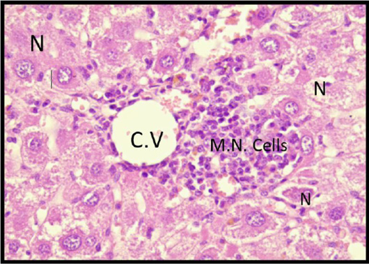
Figure 6:
Liver section of mice of C+ve group shows dilated central vein (C.V) with moderate mononuclear cell infiltration (lymphocytes and macrophages) with marked centrelobular necrosis (coagulative N.) × 40H and E stain.
Liver section of mice of C+ve group shows dilated central vein (C.V) with moderate mononuclear cell infiltration (lymphocytes and macrophages) with marked centrelobular necrosis (coagulative N.) × 40H and E stain.