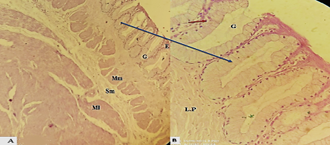
Figure 4:
Photomicrograph section illustrated the esophagus in ostrich cervical part H and E stain 100×,(B): the gland in ostrich simple tubular gland (G) (mucus gland ) PAS stain 200 ×:B: epithelium (E),( lamina propria (L.P), lamina muscularis (Mm); (Mm), tunica muscularis (MI) and (ME).