
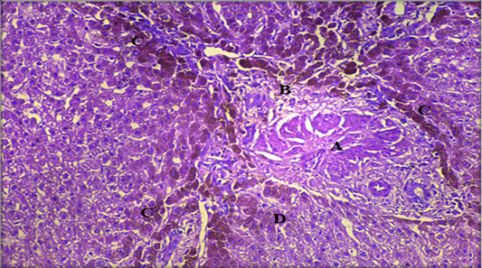
Figure 9:
Histopathological section of liver showing A: hyperplasia of bile duct which proliferation of the epithelial lining as gland, B: fibrosis of portal area with infiltration inflammatory cells, C: hemosiderin (Yellowish brown materials accumulated among hepatocytes in bile canaliculi, D: kuffper cells (H & E stain. X20).