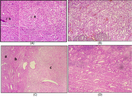
Figure 3:
A: Normal Kidney tissue (a) glomerulus (b) renal tubules 4X, B: Nephritis of Kidney with inflammatory cell aggregation in low concentration of uranium 4X. C: (a) Normal kidney tissue. (b) area of fibrosis. (c) renal cell carcinoma 4X, D: Abnormal kidney with sclerosis of glomeruli renal tubules atrophy with collection of protein material in renal tubules in low concentration of uranium 4X.
