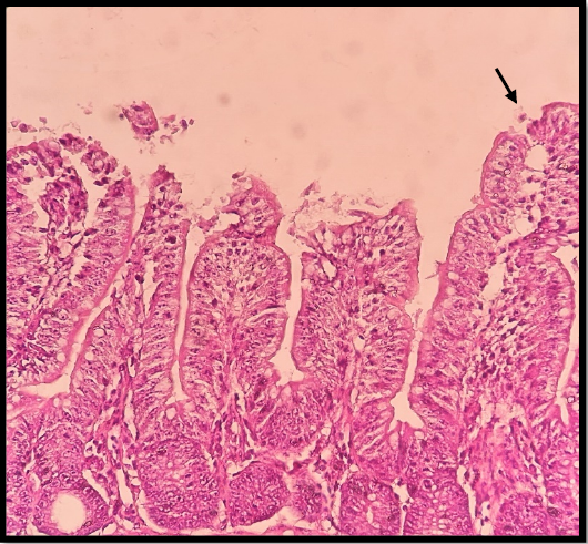
Figure 2:
Ileum lesion on G2 (8 DPI), considerable villous shortening, epithelial sloughing acceptance, and multiple established stages of parasites were visible with several Cryptosporidium oocysts attached (black arrow), H & E stain X40.