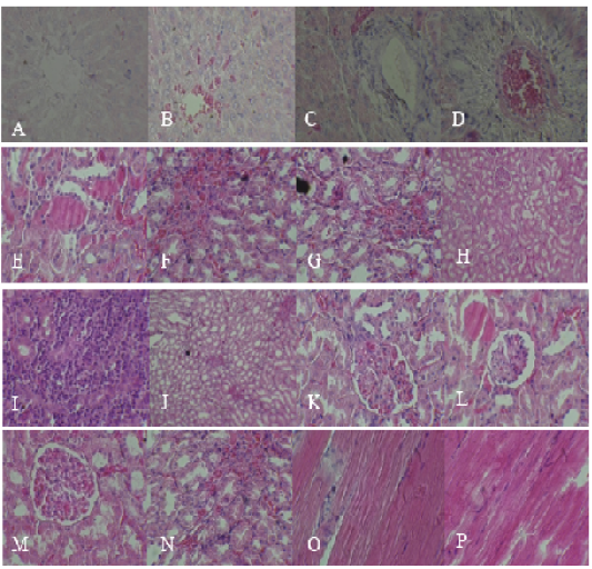
Histopathological section for different animals showing congestion of hepatic sinusoids and central vein (A), and cellular swelling (B), pericentral necrosis with infiltration of inflammatory cells (C), with fibrosis and congestion of central vein (D). The kidney sections show the presence of hyaline cast with sloughing of epithelial lining urinary tubules (E), congestion with infiltration of inflammatory cells (F, G, H and J), besides areas of necrosis (I), besides sloughing of epithelium lining urinary tubules (K), with dilatation of Bowman space (L) and congestion of the glomerular tuft with vacuolation in renal tubules (M), with degenerative changes (swelling) in the renal tubular epithelium (N). Muscle section shows infiltration of inflammatory cells besides edema causes separation of muscle’s fibers in addition to the presence of fragmentation and necrosis (O and P).