
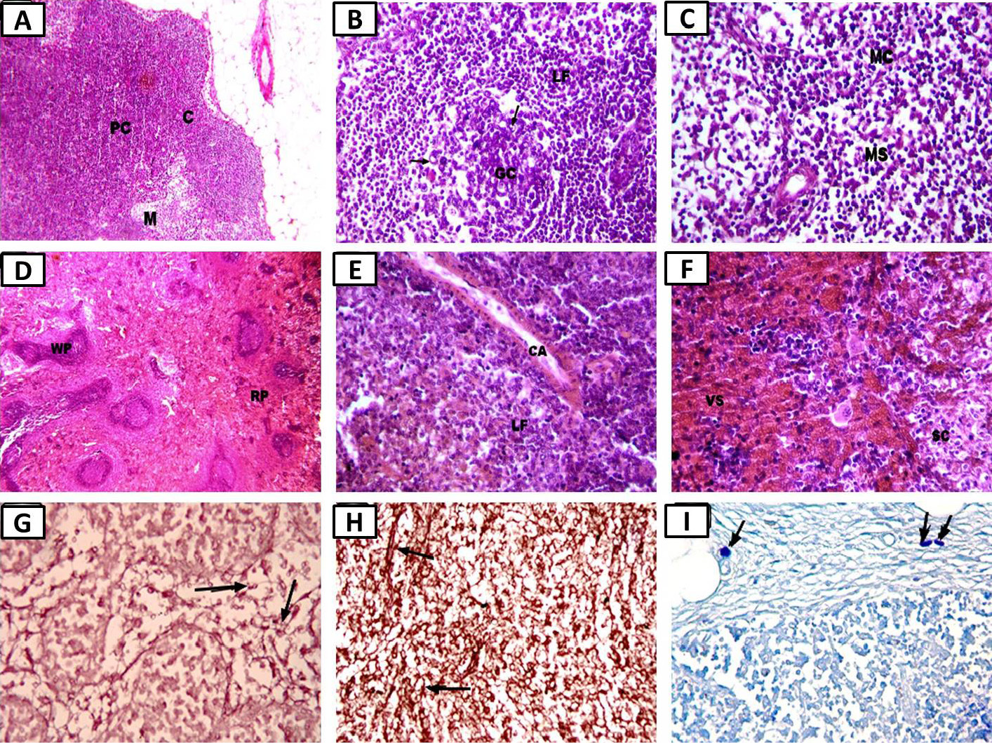
Photomicrographs of the cyclophosphamide treatment group showing: A, slight improvement in the whole structure of the lymph node (×100); B, cortical changes with some lymphocytic depletion in germinal centers and some necrotic degenerated cells (ghost cell) (arrows) (×400); C, medulla with medullary cords (MC) and medullary sinuses (MS) (×400); D, slight improvement in spleen white pulp and red pulp structure (×100); E, white pulp with dilated central arteriole (CA) in non-active lymphoid follicle (LF) (×400); F, red pulp with dilated and congested venous sinuses (VS) and splenic cords (SC) (hematoxylin and eosin, ×400); G, almost normal shape of thin, delicate elastic fibers in the lymph node (arrow) (Orcein, ×400); H, nearly normal shape of thin, elastic fibers (arrow) in the spleen (Orcein, ×400); I, almost normal mast cell size and numbers (arrows) in the pericapsular connective tissue of the lymph node (toluidine blue, ×400).