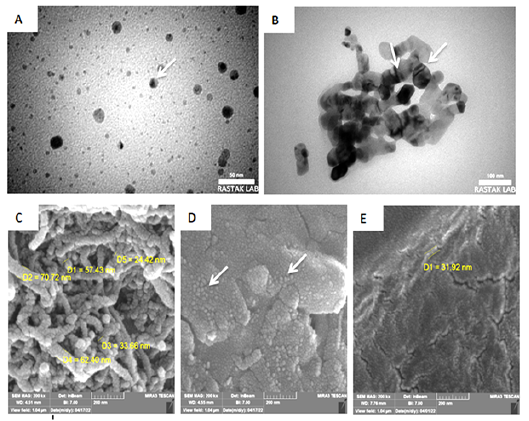
Figure 2:
Electron microscope shows scan and transmission of Nano phytosome pumpkin-lidocaine , where (A&B) are the transmission modes, the white arrows indicate multilamellar vesicles, where C, D, and D are the scan mode and the white arrows indicate spherical shapes in different sizes.