
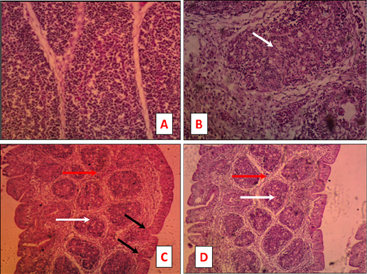
Figure 2:
Bursa tissue sections of naturally IBDV infected chicken stained by H and E. Severe depletion of the lymphoid follicle germinal center (B) deposition of interfollicular connective tissues (Fig. 1B), depletion of the lymphoid follicle tissue (C and D) accompanied with deposition of interfollicular connective tissues (C and D), and hypertrophy of the epithelial lining making finger like projection (C).