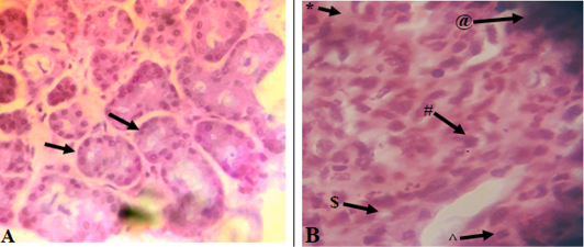
Figure 4
Microscopic images show a histological comparison between the lip tissues of an infected and a healthy sheep, where the former shows significant cellular and histological abnormalities with ulcerative and proliferative dermatitis; A: Healthy tissue with simple cuboidal epithelial forming the ducts of the salivary gland (black arrows); B: Infected tissue with a densely populated area of mononuclear phagocytic cells (personal communication with Dr. Khaled Qabaha, AAUP-Palestine) (Hash (#)), pale intracytoplasmic inclusion bodies (Caret (^)), subcorneal vesicles (Dollar Sine ($)), epithelial hyperplasia (Asterisk (*)), and necrotic crusts (At Sign (@)).